Medical Research
The Future of Surgery 3D-Printed Implants Redefining Infection Control

Researchers at Washington State University have made a new discovery: a surgical implant made of cutting-edge materials that can effectively fight bacterial infections. 3D-Printed Implants Redefining Infection Control, an innovative device, was tested in the lab and found to be able to kill 87% of the bacteria that cause staph infections. The study, which was published in the International Journal of Extreme Manufacturing, shows a possible game-changer in preventing infections during standard surgeries like hip and knee replacements that are done every day around the world. Getting bacteria on an implant is a big reason why they fail and bad things happen after surgery.
The Challenge of Infections in Implant Surgery
Amit Bandyopadhyay, a Boeing Distinguished Professor in WSU’s School of Mechanical and Materials Engineering, talked about how important it is to prevent infections during implant surgeries. He said that since implants don’t have built-in defenses against infections right now, we need a completely new way of doing things. When antibiotics are used as a preventative measure, they don’t always work. Life-threatening infections can happen right after surgery or as side effects weeks or months later.
Material Innovation

It was a big step for the WSU researchers to use 3D printing in their work, which changed the future of surgery with 3D-Printed Implants Redefining Infection Control. They made the titanium mixture used in implants better by adding 3% copper and 10% tantalum, a metal that doesn’t rust. This new combination not only had amazing antibacterial qualities, but it also worked well with the tissue around it, which could lead to a new era of implants that don’t get infections. The copper surface of the material worked especially well, breaking down bacterial cell walls when it came in touch with them. At the same time, tantalum encouraged healthy cell growth, which helped people heal faster.
Comprehensive Study and Validation

Over three years, the experts did a lot of research on their groundbreaking implant. With 3D-Printed Implants Redefining Infection Control, they are making a difference in the future of surgery. In order to do this, its mechanical qualities, biological interactions, and antibacterial effects had to be tested in the lab and on animals. The team made sure that metal ions from the implant wouldn’t move into nearby tissues and cause harm because they were worried about wear. The fact that the device could do more than one thing was a big plus; it kept infections at bay and blended in perfectly with bone tissue.
Advantages of the Multifunctional Implant
Susmita Bose, a co-author and Westinghouse Distinguished Professor talked about the multipurpose implant’s unique benefits, which change the way infection control is done. It is different from other surgical devices because it can prevent infections and help bone cells grow together. Because infections are so common in modern surgery, a gadget that is great at both of these things could completely change how well patients do.
You Might Be Like This:A New Chapter in Vision Restoration By Rebuilding Retinal Ganglion Cells
Future Prospects and Ongoing Research
The researchers are unwavering in their commitment to refining their groundbreaking implant further, shaping the future of surgery with 3D-printed Implants Redefining Infection Control. Their goals include elevating the bacterial death rate to exceed 99% without compromising tissue integration. Additionally, they aim to validate the performance of the materials under real-world loading conditions, ensuring their efficacy in scenarios that patients may encounter in their daily lives, such as hiking post-knee replacement.
Conclusion Of 3D-Printed Implants
In conclusion, the development of infection-resistant, 3D-printed implants represents a significant stride toward addressing a longstanding challenge in surgical procedures. The potential to not only control infections but also enhance the integration of implants with surrounding tissues opens new avenues for improved patient outcomes, redefining infection control. As research continues and the multifunctional implant undergoes further refinement, the prospect of a future where implant surgeries are less prone to complications and infections becomes increasingly promising. This pioneering work from Washington State University, shaping the future of surgery with 3D-printed Implants Redefining Infection Control, may very well mark the beginning of a new era in the field of surgical implants, bringing hope for safer and more effective procedures worldwide.


Game-Changer in Cancer Research: UNIST’s Nanodrones Take the Spotlight
In the realm of groundbreaking cancer treatment breakthroughs, the spotlight is now on the Ulsan National Institute of Science and Technology (UNIST), where a dynamic team of researchers has unveiled a potential game-changer. Imagine a world where tiny nanodrones, aptly named NK cell-engaging nanodrones (NKeNDs), take center stage in the fight against cancer.
RELATED ARTICLE New revolutionary nanodrones enable targeted cancer treatment

Led by the innovative minds of Professors Sebyung Kang and Sung Ho Park from the Department of Biological Sciences, this team has cracked the code to revolutionize cancer treatment. These nanodrones, far from the futuristic sci-fi portrayals, are engineered to specifically target and obliterate cancer cells, marking a significant leap forward in the battle against this relentless disease.

At the heart of this breakthrough lies the ability of these nanodrones to engage natural killer (NK) cells, the body’s frontline soldiers against cancer. What sets the NKeNDs apart is their precision – think of them as nanoscale guided missiles homing in on cancer cells with unparalleled accuracy. The secret sauce involves utilizing AaLS protein cage nanoparticles as the foundation for these nanodrones, incorporating specific cancer-targeting and NK cell-recruiting ligands, aptly named HER2 @NKeND and EGFR@NKeND.
Lab tests have showcased the remarkable ability of these nanodrones to selectively bind to various types of cancer cells while rallying NK cells to mount a defense against the invaders. The real breakthrough emerged during mice trials, where administering HER2 @NKeNDs alongside human immune cells resulted in a significant slowdown in tumor growth, all without adverse effects.

Professor Kang Se-byung, brimming with excitement, highlighted the potential for customizing treatments for different cancers using these NK cell delivery nanodrones. It’s not merely about targeting cancer cells; it’s about doing so with surgical precision, minimizing collateral damage and maximizing the impact of the body’s immune system.
MORE LATEST ARTICLE Brain Clot Revolution, Vortex Ultrasound Tornado in Brain Health
This groundbreaking study, published in Nano Today, marks a pivotal moment in scientific progress. With the support of various institutions dedicated to advancing knowledge, the door to a new era in cancer treatment swings wide open. Nanodrones may just be the superheroes we’ve been yearning for, offering hope and resilience in the face of one of humanity’s most formidable adversaries. As we raise a toast to science and innovation, the journey towards conquering cancer takes a remarkable leap forward. Cheers to the heroes of the microscopic world.
Medical Sciences
Revolutionizing Hypertension Treatment, Zilebesiran’s Promise in Cardiovascular Health

Zilebesiran’s Pioneering Role in Hypertension Care
In the dynamic realm of cardiovascular research in 2023, the American Heart Association (AHA) has spotlighted groundbreaking advances that promise to reshape the landscape of heart health. Researchers have delved into various dimensions, from innovative drug therapies to cutting-edge procedures, offering hope for improved prevention and treatment of cardiovascular diseases.
RELATED ARTICLE AHA names top advances in cardiovascular disease research for 2023

Brief Introduction about zilebesiran
A solitary administration of the experimental drug zilebesiran demonstrated both safety and efficacy in reducing systolic blood pressure in individuals with mild-to-moderate high blood pressure for a duration of up to six months. This information, disclosed in the Phase 2 segment of the KARDIA study, was presented as late-breaking scientific findings during the American Heart Association’s Scientific Sessions 2023. The conference, held from November 11 to 13 in Philadelphia, serves as a leading global platform for the exchange of cutting-edge scientific developments, research insights, and updates on evidence-based clinical practices in the field of cardiovascular science.
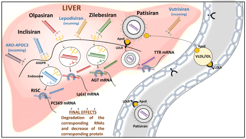
Zilebesiran functions as an investigational RNA interference agent designed to target angiotensinogen (AGT), a hormone primarily produced in the liver that plays a significant role in the regulation of blood pressure.
RELATED INFORMATION New drug zilebesiran effectively lowers blood pressure for six months, study finds
One notable stride involves a novel drug targeting hypertension. In this arena, Zilebesiran, an investigational drug, has emerged as a potential game-changer. In a phase I study involving 107 participants aged 65 and older with high blood pressure, the drug exhibited promise by reducing blood pressure measurements over an eight-week period. The study demonstrated efficacy, even with a single dose, offering potential advantages in terms of accessibility to care and adherence to long-term drug regimens. Moreover, the drug’s impact extended beyond hypertension, sparking excitement about its potential therapeutic applications for kidney and heart diseases.
Another significant breakthrough lies in the realm of stroke treatment. Endovascular thrombectomy, a minimally invasive procedure to remove blood clots causing strokes, has traditionally been applied to medium-sized strokes. However, new studies from various regions, including China, North America, Europe, Australia, and New Zealand, have presented compelling evidence that this procedure could benefit individuals with larger, more severe strokes. The trials, published in renowned journals such as the New England Journal of Medicine and The Lancet, underscored the procedure’s superiority in improving functional independence and reducing disabilities in severe stroke patients.

Advancements in imaging techniques have also taken center stage, particularly in guiding stent placement for individuals with complex coronary lesions. While angiography remains the standard for guiding stent placement, intravascular imaging, including techniques like optical coherence tomography (OCT), has showcased its potential. Studies such as ILUMIEN IV and OCTOBER compared OCT-guided percutaneous coronary intervention (PCI) to angiography-guided PCI, revealing that OCT guidance resulted in a larger minimum stent area, suggesting improved outcomes in complex cases.
The intersection of atrial fibrillation (AFib) and stroke treatment has seen intriguing developments. A study published in the New England Journal of Medicine challenges the conventional timeline for initiating anticoagulant treatment in AFib patients following a stroke. Contrary to current practice guidelines, early initiation of Direct-Acting Oral Anticoagulants (DOACs) within 48 hours of a minor or moderate stroke, and on day six or seven following a major stroke, demonstrated promising results. The study suggests that CT or MRI scans could offer more precise insights into stroke severity, aiding in identifying individuals who could benefit from earlier DOAC use.
Furthermore, the realm of cardiovascular health has expanded its reach to individuals without diabetes. Semaglutide, a medication primarily approved for long-term weight management in individuals with obesity, has exhibited potential cardiovascular benefits. Studies indicated that it not only lowered the risk of heart problems in people with diabetes but also showcased effectiveness in reducing heart failure-related symptoms and cardiovascular-related death in overweight and obese individuals without diabetes.

The AHA has also emphasized the role of social determinants of health in cardiovascular disparities. Research linking cardiovascular death rates to income status, access to healthy foods, and housing highlights the need to address these social factors for effective cardiovascular disease prevention and management.
On a broader scale, the AHA has taken a holistic approach with the introduction of the cardiovascular-kidney-metabolic (CKM) syndrome. This syndrome, encompassing the intricate interconnections between obesity, chronic kidney disease, diabetes, and cardiovascular disease, has prompted the development of a new risk calculator. The American Heart Association PREVENT risk calculator now estimates 10- and 30-year risks for heart attacks, strokes, and heart failure, incorporating measures of cardiovascular, kidney, and metabolic health, along with considerations for social determinants of health.
READ MORE SCIENTIFIC ARTICLE Brain Clot Revolution, Vortex Ultrasound Tornado in Brain Health
Lastly, the spotlight extends to the treatment of peripheral arterial disease (PAD), particularly chronic limb-threatening ischemia. Studies such as BASIL-2 and BEST-CLI have compared the effectiveness of surgical bypass to endovascular therapies, offering insights into optimal intervention choices for improved outcomes.
In essence, the cardiovascular research landscape of 2023 paints a vivid picture of innovation, ranging from novel drug therapies to sophisticated procedures and holistic risk assessment tools. As we navigate this evolving landscape, the promise of enhanced prevention, treatment, and overall cardiovascular health stands as a beacon of hope for individuals worldwide.
Medical Sciences
Decoding Waardenburg Syndrome,Genetic Aspects and Clinical Nuances of Waardenburg Syndrome

Understanding Waardenburg Syndrome: Genetic Aspects and Clinical Insights
Introduction
Waardenburg syndrome stands as a rare genetic disorder renowned for its distinctive features involving sensorineural hearing loss and pigmentary abnormalities affecting the hair, skin, and eyes. Named after the Dutch ophthalmologist Petrus Johannes Waardenburg, who first observed the correlation between deafness and heterochromia iridis, this syndrome has been a subject of keen medical interest.
RESOURCED ARTICLE Waardenburg syndrome

Demographics and Frequency: Waardenburg syndrome does not discriminate; it affects both males and females equally and can manifest across all races. With a population frequency of approximately 1 in 40,000, it contributes to 2–5% of all cases of congenital deafness.
Genetic Basis: This neurocristopathy arises from gene mutations affecting neural crest differentiation during embryonic development. Various genes play pivotal roles, and their mutations give rise to different types of Waardenburg syndrome. Noteworthy genes include:
- PAX3 (Chromosome 2q): Implicated in WS1 and WS3.
- MITF (Chromosome 3p): Associated with WS2A.
- Chromosomes 1p, 8p, 8q, 13q, 20q, and 22q: Linked to WS2B, WS2C, WS2D, WS4A, WS4B, and WS4C, respectively.
The mutations encompass various types such as insertions, deletions, frameshifts, splice alterations, missense, or nonsense mutations. Interestingly, most types of Waardenburg syndrome follow an autosomal dominant inheritance pattern, meaning that one affected gene passed on to a child is sufficient for them to exhibit the syndrome. However, complexities arise with genes like EDN3 or EDNRB, which generally follow autosomal recessive patterns.

Clinical Features: Waardenburg syndrome manifests early in life, and its diagnosis can be challenging due to the subtlety of clinical signs. Major types (Type 1-4) display distinct features:
- Type 1: Most common, characterized by sensorineural deafness, distinct facial features, white skin patches, and pigmentary abnormalities in the eyes.
- Type 2: Similar to Type 1 but lacks abnormalities in the inner canthi.
- Type 3 (Klein-Waardenburg syndrome): Includes musculoskeletal abnormalities.
- Type 4 (Shah-Waardenburg syndrome): Similar to Type 2 but with the addition of Hirschsprung syndrome.
Diagnosis: The diagnosis of Waardenburg syndrome relies on clinical features, with major criteria encompassing sensorineural deafness, iris pigmentary abnormalities, and hair pigmentation anomalies. Minor criteria include features like unpigmented skin patches, synophrys, and premature greying of scalp hair. The W index, calculated based on canthal distances, aids in identifying dystopia canthorum. Genetic sequencing, particularly of the PAX3 gene, can provide a definitive diagnosis and guide genetic counseling.

Treatment and Management: As of now, there is no curative treatment for Waardenburg syndrome due to its genetic nature. Genetic counseling is crucial for affected individuals contemplating starting a family. Regular audiology examinations are recommended for managing deafness, and those with Hirschsprung syndrome may require surgery. Sun protection is emphasized for areas with unpigmented skin patches.
Outlook: Waardenburg syndrome presents stable clinical features throughout life, and individuals can expect a normal life expectancy. Although the syndrome poses challenges, advancements in genetic understanding have paved the way for improved diagnostics and counseling, offering individuals and families affected by Waardenburg syndrome a clearer path forward.
READ MORE INTERESTING ARTICLE Cortex Chronicles, Paul G. Allen’s Institute Explores the Study on Visual Masking in Bioscience
In conclusion, the intricate genetic landscape of Waardenburg syndrome underscores the importance of ongoing research and medical advancements in unraveling the complexities of this rare disorder. As we delve deeper into the genetic intricacies, our ability to diagnose, manage, and provide support for individuals with Waardenburg syndrome continues to evolve.
-
 Medical Sciences8 months ago
Selenium Nanoparticles Redefining Postmenopausal Osteoporosis Treatment with a Novel Approach
-

 Medical Sciences8 months ago
Transforming Heart Failure Care: Unveiling Abbott’s ARIES Trial Breakthrough with Aspirin-Free HeartMate 3
-

 Medical Sciences7 months ago
Nanodrones Against Cancer,UNIST’s Innovation Marks a New Era in Treatment
-

 Medical Sciences8 months ago
Geriatric Care in the Face of Non-ST Elevated Myocardial Infarction and Acute Coronary Syndrome
-

 Blog8 months ago
UK’s Milestone In Genetic Medicine,CRISPR Therapy Treating Sickle-Cell Disease and β-Thalassaemia
-

 Medical Research8 months ago
A New Chapter in Vision Restoration By Rebuilding Retinal Ganglion Cells
-

 Blog8 months ago
Cutting-Edge Diagnostic Technology Handheld Device for Alzheimer’s and Parkinson’s Diseases
-

 Medical Sciences8 months ago
Regenerative Surgery A Comprehensive Innovation by Stem Cells in Face and Eye Transplantation